- Today
Visualisation of reticular veins when treating telangiectasia
- Zola Mzimba
- Medical Technology
Published: 13th November 2026
Author: Zola Mzimba, Venous Specialist
TL;DR (Key Clinical Takeaways)
Target the Root Cause: Treating visible spider veins (telangiectasia) alone often leads to disappointing results; you must identify and treat the underlying, hidden "feeding" reticular veins first to achieve lasting cosmetic success.
Tech Overcomes Blind Spots: Feeding veins are frequently invisible to the naked eye, especially in higher Fitzpatrick skin types, making specialised visualisation devices essential for accurate clinical evaluation.
Match the Tool to the Task: While affordable optical tools like transillumination map superficial paths, advanced high-frequency ultrasound (18–22 MHz) is unmatched for measuring depth, tracking flow and providing real-time needle guidance during cannulation.
Visualising Feeding Reticular Veins: Why It Matters and Which Devices Can Help
Why Is It Important?
Patients frequently present with cosmetically unsatisfactory telangiectasia (spider veins), which they wish to have treated. However, closer examination often reveals underlying reticular veins feeding these clusters.
Telangiectasia are small intradermal vessels measuring less than 1 mm in diameter, whereas reticular veins are larger subdermal vessels measuring 1–3 mm. Despite their deeper location, reticular veins may still be visible through the skin.
Identifying these feeding veins is important because treating superficial telangiectasia alone may produce disappointing results. Where a feeding relationship is demonstrated, addressing the reticular veins first may improve cosmetic outcomes and reduce the need for additional treatments.
Clinical Examination
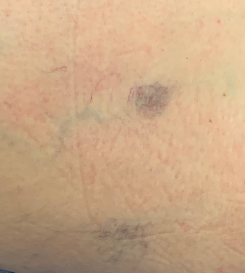
Picture 1 – Telangiectasia with associated feeding reticular veins.
The diagnostic distinction between telangiectasia and reticular veins is based primarily on vessel diameter and anatomical location.
Both may be visible to the naked eye, but visibility depends on vessel depth, skin translucency, pigmentation and lighting. Reticular veins are generally easier to identify in Fitzpatrick skin types I and II than in higher skin types.
Importantly, feeding reticular veins may be present despite being invisible to the naked eye. Several visualisation devices can assist their identification, ranging from simple optical systems to high-frequency ultrasound.
Transillumination Devices (Veinlite)
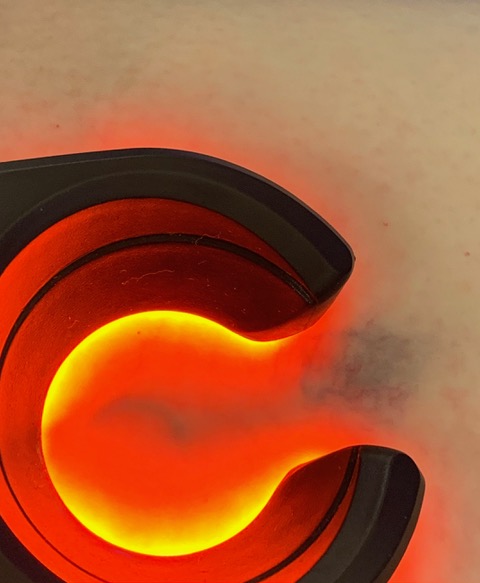
Picture 2 – Transillumination demonstrating feeding reticular veins.
Transillumination devices direct light into the skin and surrounding tissues. Blood absorbs the light, making veins appear dark against the illuminated background.
This enhances visualisation of reticular veins that may otherwise be difficult to identify, particularly in patients with higher Fitzpatrick skin types. Depending on the device and tissue characteristics, vessels several millimetres beneath the skin may be visualised.
Importantly, transillumination helps identify feeding reticular veins, allowing more accurate targeting during sclerotherapy. Treating these vessels before superficial telangiectasia may improve cosmetic outcomes.
These devices are relatively inexpensive, with some models costing a few hundred pounds.
Near-Infrared Vein Visualisation Devices
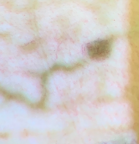
Picture 3 – Near-infrared imaging demonstrating the course of subdermal veins.
These devices use near-infrared light, which penetrates deeper into tissues than visible light. Haemoglobin absorbs near-infrared light, allowing sensors to detect underlying veins and project their image onto the skin surface.
Depending on the device and tissue characteristics, veins may be visualised at depths of approximately 5–10 mm, making these systems useful for identifying feeding reticular veins that may not be visible to the naked eye.
By demonstrating the location and course of these vessels, near-infrared devices can assist targeting during sclerotherapy.
However, they cannot accurately determine vessel depth or assess venous reflux. Image quality may also be affected by tissue thickness, pigmentation and scarring.
These systems typically cost several thousand pounds.
Cross-Polarised Light Devices
Cross-polarised light devices use polarising filters to reduce light reflected from the skin surface, improving visualisation of superficial dermal vessels.
This technique provides excellent visualisation of telangiectasia by reducing surface glare and enhancing vascular contrast.
However, its ability to demonstrate deeper feeding reticular veins is limited. Although superficial vessels may be clearly demonstrated, reticular veins lying further beneath the dermis may remain invisible.
Cross-polarisation is therefore particularly useful for assessing and documenting telangiectasia but is less reliable for identifying deeper feeding reticular veins.
High-Frequency Ultrasound
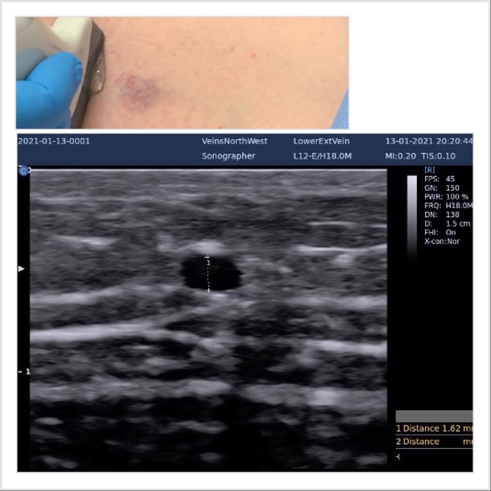
Picture 4 – High-frequency ultrasound demonstrating a reticular vein and needle placement.
Conventional linear ultrasound probes used for varicose veins can visualise reticular veins measuring 1–3 mm. However, identifying a vessel is different from accurately guiding a needle into its lumen.
Although a 10 MHz probe can resolve structures considerably smaller than 1 mm, reliable needle placement requires sufficient resolution to distinguish the vessel walls, lumen and needle tip.
Higher-frequency probes, typically 18–22 MHz, provide improved visualisation of superficial structures, making them particularly useful for ultrasound-guided reticular vein sclerotherapy.
Unlike optical devices, ultrasound can assess vessel depth, diameter, course and blood flow. However, higher frequencies have reduced tissue penetration, and successful cannulation depends on probe quality, operator experience and needle visibility.
These frequencies are therefore advantageous rather than universally essential.
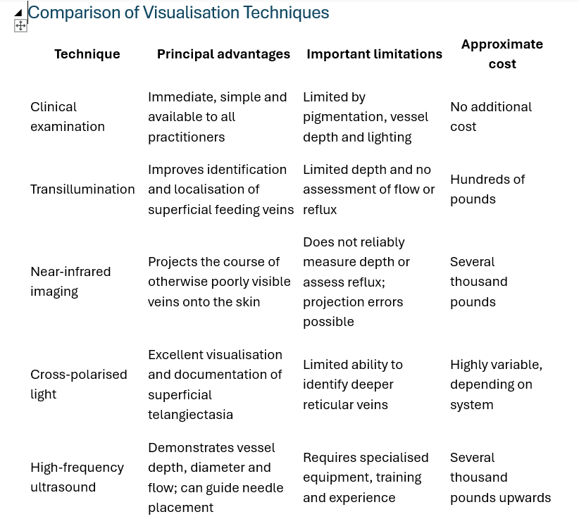
Clinical Implications
There are three distinct challenges when treating feeding reticular veins:
1. Visualisation: Identifying the presence of a feeding reticular vein.
2. Localisation: Establishing its course and, where possible, its depth beneath the skin.
3. Cannulation: Accurately placing a needle within the vessel lumen to deliver treatment.
Different technologies address these challenges to varying degrees.
Clinical examination and optical devices can improve identification and localisation, but they cannot reliably determine vessel depth or confirm intraluminal needle placement.
High-frequency ultrasound offers additional anatomical information and real-time needle guidance, although it is not required for every reticular vein treatment.
Conclusion
Effective treatment of telangiectasia requires recognition of any underlying feeding reticular veins, which may not always be visible on clinical examination.
Several techniques are available to improve their visualisation, ranging from simple transillumination devices to more sophisticated near-infrared imaging and high-frequency ultrasound. Each has advantages and limitations, particularly regarding vessel depth, localisation and needle guidance.
The choice of technology should reflect the clinical requirements and available resources.
Ultimately, the aim is not simply to treat the visible telangiectasia, but to identify and address any associated feeding reticular veins that may influence the final cosmetic outcome.
Summary
About the author

Zola Mzimba, Venous Specialist
Zola Mzimba is a venous specialist based in Northern Ireland. Initially trained as a vascular surgeon for the last decade in both the NHS and the independent sector, he has restricted his practice to and specialises solely in venous disease. He was the co-chair involved in centralising varicose vein services to two high throughput centres in Northern Ireland. He is based in Derry and consults at Jace Medical Clinic, as well as for the VeinCentre group in Belfast. He is a registered venous sonographer and also has the Diploma in Phlebology from the European College of Phlebologists.